Introduction
Space closure often poses challenge to orthodontist. Orthodontic tooth movement has always been limited to action and reaction reciprocal effects in terms of anchorage.1 Individual canine retraction creates space distal to incisors which is esthetically unpleasant and also increases treatment duration. En-masse retraction requires higher amount of forces and creates reciprocal effects and taxes molars in terms of anchorage.
The versatility of Mini-screw implants (MSIs) has increased its demand in orthodontics. They reduce patient compliance, provide absolute anchorage, minimize reciprocal effects and thus reduce overall treatment duration. Because of their advantageous effects they can be used for various purposes as like for absolute intrusion, en-masse retraction, molar distalization etc.2 Bi-alveolar dental protrusion is one of the common malocclusion encountered by orthodontist and the most common treatment modality includes extraction of 4 first premolars followed by intrusion and retraction of maxillary anteriors to obtain ideal dental and soft tissue profile.3 Prolonged duration of treatment and stress generated because of force application are two important factors responsible for orthodontically induced root resorption. On force application to a tooth, initial tooth displacement is produced and then orthodontic tooth movement starts. Thus it is utmost important to study the amount of stress produced and pattern of stress distribution in PDL after force application.
Finite Element Methodology is a highly precise technique used in engineering to analyze structural stress on the basis of physical properties of structure being analyzed. The first descriptive Finite Element Analysis ( FEA) study of orthodontic tooth displacement and stress magnitude was conducted by Tanne et al.4 Over the years finite element method has been used successfully to simulate various orthodontic tooth movement and analyze structural stress. In the present study FEM model is used to evaluate stress distribution pattern in upper anterior region during simultaneous intrusion and retraction of upper six anterior teeth using bilateral posterior implant and one anterior mid-implant.
Materials and Methods
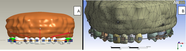
Finite Element Model was generated using ANSYS 8.1 workbench 11 software which was replica of human maxilla. It consisted of periodontal ligament, alveolar bone with two bilateral posterior and on anterior mid mini-implant along with all maxillary teeth except first premolar bilaterally. MBT .022 bracket system was simulated and the arch wire used was 0.019”X0.025” stainless steel consisting of power arm distal to lateral incisors bilaterally and a v-bend in the centre between central incisors. (See Figure 1 a: and b)
Various steps involved in Finite Element Model generation was followed accordingly described by Sagar et al.5, 6
Construction of geometric model.
Conversion of the geometric model to a finite element model.
Material property data representation.
Defining boundary condition.
Loading configuration.
Construction of geometric model: In this study a 3D CT scan of maxilla was taken including all maxillary teeth except maxillary first premolar bilaterally. Mathematical model produced represented the biological properties of the teeth and the periodontium. This was represented in terms of points(grids), lines, surface(pattern) and volume (hyperpatches). The software used for geometric modeling was ANSYS workbench 11.
Conversion of the geometric model to a finite element model: The geometric model was converted into finite element model. The finite element modeling is the representative of geometry in terms of finite number of elements and nodes. This process is called discretization. The main idea behind discretization is to improve the accuracy of the result. The elements are interconnected at points which are called as nodes. (Table 6)
Material property data representation: The different structures involved in this study includes teeth, the periodontal ligament and the alveolar bone. Each structure has specific material property. The material properties used here was derived by Mc Guiness and were also used in finite element studies done by Tanne et al. These materials properties were the average values reported in literature.7 (Table 7).
Defining boundary condition: The boundary condition were defined to simulate how the model was constrained and to prevent it from free body motion. The nodes attached to the corner and outside surface of the bone are fixed in all direction, to avoid free body movement of the tooth.
Loading configuration: An Intrusion force of 120gm was applied with the help of nickel titanium coil springs between maxillary central incisors through a mid mini implant placed at 11mm from interdental papillae in the centre and retraction forces of 200gm were applied simultaneously with the help of nickel titanium coil springs from posterior implants bilaterally, placed at 8mm from interdental papillae between maxillary second pre-molar and first molar to the power arm at 5.9mm of height between canine and lateral incisor. Stress generated were observed around maxillary anterior teeth, alveolar bone and around implants bilaterally.( See Figure 2)

Results
The stress values on teeth were found to be not significantly different when compared to left side with right side. (See Figure 3 a & b, Figure 4, Figure 5)
Table 0
|
Stress on teeth |
|
|---|---|
|
Right Central=0.106(Mpa) |
Left Central=0.105(Mpa) |
|
Right Lateral=0.410(Mpa) |
Left Lateral= 0.390(Mpa) |
|
Right Canine=0.062(Mpa) |
Left Canine=0.058(Mpa) |
Table 0
|
Soft bone |
|
|---|---|
|
Right Central=9.85E-03(Mpa) |
Left Central=1.08E-02(Mpa) |
|
Right Lateral=1.01E-02(Mpa) |
Left Lateral= 1.01E-02(Mpa) |
|
RightCanine=1.33E-02(Mpa) |
Left Canine=1.34E-02(Mpa) |
Table 0
|
Hard bone |
|
|---|---|
|
Right Central=9.98E-02(Mpa) |
Left Central=1.01E-01(Mpa) |
|
Right Lateral=9.56E-02(Mpa) |
Left Lateral=1.02E-01(Mpa) |
|
Right Canine=1.01E-01(Mpa) |
Left Canine=1.02E-01(Mpa) |
Table 0
|
Tooth deformation |
|
|---|---|
|
Right Central=6.528E-06(Mpa) |
Left Central=6.439E-06(Mpa) |
|
Right Lateral=6.034E-06(Mpa) |
Left Lateral= 6.049E-06(Mpa) |
|
Right Canine=3.565E-06(Mpa) |
Left Canine=3.995E-06(Mpa) |


Discussion
The perception of esthetic has changed with time and it differs in different places in different ethnic groups around the world. The maxillary anterior segment forms a very important part of facial esthetics. One of the most common malocclusion related to upper anteriors is bi-alveolar dental protrusion characterized by increased overjet and overbite. Improving esthetic by correction of incisor relationship is prime and foremost goal of an orthodontist. In orthodontics, the study of initial stress concentration in the periodontal ligament, teeth and surrounding structures is important because ligament act as a mediator for the tooth movement and any alteration from optimal force produces adverse effect on the tooth and the periodontium as apical root resorption and alveolar bone loss. It has been reported that the bodily movement of the anterior teeth can be achieved by directing the force through the centre of resistance of anterior teeth, by altering the occluso-gingival location of mini-implants and length of the anterior retraction hook (ARH).7 The demand of speedy and efficient orthodontic treatment has been increasing in recent years. To meet this demand, sliding mechanics in combination with implant anchorage has become widely popular throughout the world. However, mini-Implant, have proven to be a useful addition to the orthodontist armamentarium for control, of anchorage and has gained enormous credibility in the clinical management of various orthodontic tooth movement.8, 9 Mini screws and power arm in the present study were placed according to the studies done by Shrinivas et all and Heydayati et all who suggested posterior implant placement at 8-10mm with power arm of 5-6mm length between canine and lateral incisor to obtain bodily translation of teeth during en-masse retraction of anterior teeth.10, 11 In the present study mini implants were incorporated to preserve anchorage during simultaneous intrusion and retraction of maxillary anterior teeth and stress generated was evaluated via FEM.
FEM a highly precise technique to calculate stress on different structures was used to analyze stress on maxillary anterior teeth. When the model was analyzed for stress generation on teeth, it was observed that the stresses around lateral incisors were found to be higher side when compared with the central incisor and canine. The stress value were found to be not significantly different when compared to left side with right side. Less pericemental area and closer to the oblique retraction force due to attachment of lever arm, might have resulted in more stress concentration around lateral incisors. Our findings are well supported by the study done by Eric J Liou who reported higher amount of root resorption on lateral incisors than other teeth during en-masse retraction and intrusion of maxillary anterior teeth with mini screws.12 In the present study maximum stress in periodontal ligament was found in cervical area than in apical region, however stress nature changes from tensile to compressive from cervical area to root apex. Studies done by Parag Bohra et all revealed the same result as accordance to present study.13 In present study, it was found that under all conditions, maximum stresses were concentrated at the cervical portion of the PDL. This is in agreement with study by S. Rex et all and Jayan Bharath et all which concluded that intrusion, extrusion, and rotational forces produces more stress at apex, whereas bodily movement and tipping forces concentrates more at alveolar crest.14, 15
When stress around the bone was analyzed it was observed that maximum stress were found around canines bilaterally in both hard and soft bone than the central and lateral incisors. As the canine at the back barred most of the retraction force, it causes distal crown tipping of canines in the extraction space. As intrusive force from the mid mini-implant was far from canine, it has less control on distal tipping of canine and the torque transmitted to maxillary canine could be insufficient to counteract distal crown tipping. FEM study done by Ahmed Othman et all supports the findings of present study where they have found that maximum stress concentration was around canine during space closure by different mechanics.16 Our study results were correlated with the finding observed in studies undertaken by Laljit et al.17, where en masse retraction of maxillary anterior teeth with the help of implants generates maximum stress at the head of implant.
Conventional en mass retraction produces extrusion of upper anterior teeth, thus the application to patient with vertical growth or deep bite or gummy smile may cause unfavorable results. Thus the FEM model generated in our study with 3 implant system i.e. two bilateral posterior implants and one mid implant between centrals was according to the study done by Monica Nimburi,18 which shows that the 3 implant system produces more bodily movement with no labial flaring of upper anterior teeth. Single implant in the anterior region produces less discomfort to the patient as well as also compensates for the curves and bends that are given during conventional retraction to overcome the side effects like deepening of bite by applying additional vertical component of intrusive forces.
The present study shows significantly lesser amount of stress in apical region compared to cervical areas as analysis shows the stresses generated at the initial time of force application and not over a period of time. Thus time dependent reaction is still unpredictable and requires more clinical evidence.
Conclusion
Intrusion and retraction of maxillary anteriors are two major phases of orthodontic treatment. Anchorage preservation should be the key factor to minimize undesirable effects. The forces should be closer to the centre of resistance to obtain bodily movements. Addition intrusion force along with the retraction force have better control over lingual crown displacement and true intrusion can be obtained. The single FEM model in the present study provides information related to every component of the periodontium along with the mini-implants. The stress value obtained in the present study is comparatively low and are more uniformly distributed.
Thus the present model simulation with one single anterior and two posterior implant can be considered as most reliable condition to obtain controlled intrusion and retraction of maxillary anterior teeth. This FEM model has demonstrated that such an approach can be valued in detailed study of orthodontic biomechanics.