Introduction
Aesthetics, as defined by Merriam-Webster dictionary, is “a branch of philosophy dealing with the nature of beauty, art and taste and with the creation and appreciation of beauty”. Aesthetics is central to dentistry when it comes to building a pleasant smile that boosts the individual’s confidence and improves their quality of life.1 Smile and facial attractiveness are strongly correlated. The present emphasis on the soft tissue paradigm is towards enhancing facial aesthetics and creating a beautiful smile. Patients today judge their treatment outcomes not only by the occlusion and alignment but also by the smile aesthetics, making it a primary reason why they seek orthodontic treatment.
The degree of gingival display and smile arc have an impact on the aesthetics of a smile. It has been said that a smile with a minimal gingival display is more aesthetic than one with a large gingival display. The buccal corridor runs from the lip commissure to the buccal surface of the most prominent maxillary posterior teeth. Nowadays, a wide smile with reduced buccal corridor is thought to represent youth and health. Tooth extractions are a common form of treatment in Orthodontics. It has been suggested that orthodontic treatment may cause a narrow maxillary arch, especially when maxillary premolars have been extracted, and that this dark area, together with a flat profile, can lessen facial attractiveness. 2 Contradictory results were found by Johnson and Smith, 3 but they all led to the same conclusion: there was no correlation between extraction aesthetics and features related to the buccal corridor or other measurements of the relationship between the width of the dentition and the mouth when smiling.
The objective of the present study was to evaluate the effect of extraction versus non-extraction treatment on the smile aesthetics of patients with Angle’s Class I malocclusion, with an objective evaluation using smile variable measurements. This study assumes added importance in that there are very few studies highlighting and comparing the effect of first and second premolar extraction on facial aesthetics.
Materials and Methods
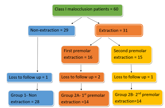
The study was conducted in accordance with the Declaration of Helsinki and had been approved by the Institutional Ethical Committee (IEC/MES/54/2020 dated 05/01/2021). The sample size was calculated for a Type I error rate of 5% and a power of 80% and comprised 60 subjects of age ranging from 12 to 25 years. The subjects were split into two groups, non-extraction (Group 1=29) and extraction groups -Group 2=31. The extraction group was further divided into first premolar (Group 2A=16) and second premolar (Group 2B=15) extraction groups. The study sample was selected after fulfilling the requirements for inclusion and exclusion criteria and included pre- and post-treatment facial frontal photographs and study models of subjects who had undergone fixed orthodontic treatment with or without four premolar extractions in patients with Angle’s Class I malocclusion. Informed consent/ assent was obtained before the photographs of the subjects were taken. At the end of the study period, loss to follow-up resulted in the inclusion of 28 patients in Group 1 and 14 patients each in Group 2A and Group 2B. (Figure 1)
Evaluation of miniaesthetics
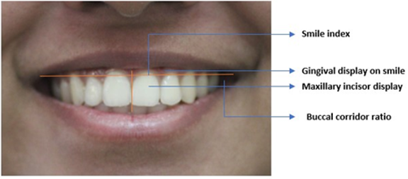
Photographic evaluation was recorded in static images with a posed smile and in natural head position (NHP). The CANON 1500 DSLR camera, which was set up on a tripod stand at a fixed distance of 20 inches, was used to take all pictures in the same setting and with the same lighting conditions. The patient was asked to say "cheese" and then smile. The images were then imported into computer software (Adobe Photoshop 7.0.1) and cropped with vertical limitations to display the immediate perioral area (drawn from the zygomatic prominence, the tip of the nose, and the soft tissue pogonion). The resolution of each image was changed to 1200 by 761 pixels. For this study, measurements were taken using the ruler tool in the Adobe Photoshop software. The following smile parameters were assessed: gingival display, smile index, maxillary incisor display on smile and buccal corridor ratio (Figure 2) (Table 1).
Statistical analysis
The SPSS statistical software (Version 22-SPSS Statistics for Windows, IBM Corp., Armonk, NY, USA) was used for the statistical study. The significance threshold was established at p<0.05. To evaluate the mean and standard deviation of the various groups, descriptive statistics were used. Normality of the data was assessed using Shapiro-Wilk test. T test was used in inferential statistics to determine the difference between time intervals. One-Way ANOVA was used to analyse data from three groups, and Tukey's HSD post hoc analysis was used to determine the difference between any two groups. Intra- examiner reliability with intra-class coefficient ranging from 0.94-0.97 suggesting reliability in intra-examiner measurements.
Table 1
Smile parameters
|
Parameter |
Definition |
|
Maxillary incisor display on smile |
Amount of vertical display of the maxillary central incisors was measured in mm. |
|
Gingival display on smile |
Normally, the display of teeth and gingival line is about 1mm or just above the cervical margins of teeth in posed smile. High smile line/ average smile line/ low smile line. |
|
Smile index |
Described by Ackerman et al. 2 (1998) as: Smile index= Inter commissural width/Interlabial gap. The lower the smile index, the less youthful the smile appears. |
|
Buccal corridor ratio |
The buccal corridor ratio was calculated according to the method given by Frush and Fisher12 (1958) as: Buccal corridor ratio(%)=Inter commissure width Visible maxillary dentition width /Inner commissure width |
Table 2
Maxillary incisor display, gingival display on smile
Table 3
Smile index, Buccal corridor ratio
Results
The present study was done to assess the miniaesthetics and arch width changes in subjects with Angle’s Class I malocclusion who underwent Orthodontic treatment with or without extraction of four premolars. Miniaesthetics parameters in this study included maxillary incisor display on smiling, gingival display on smiling, smile index and buccal corridor ratio. Arch width measurements of intercanine, interpremolar and intermolar widths were done using digital vernier caliper.
Maxillary incisor display- While analysing the maxillary incisor display on smiling, it was found that there was an increase in maxillary incisor display in the non-extraction group (Group 1- 1.39±0.34) while first premolar extraction group (Group 2A) showed a decrease (2.31±0.50). Second premolar extraction group (Group 2B) showed no significant change in maxillary incisor display (0.90±0.19). (Table 2)
Gingival display on smiling- Comparison of pre- and post-treatment gingival display on smiling in all the 3 groups showed no significant change with a mean difference of 0.07±0.07mm, 0.38±0.65mm, 0.07±0.15mm in non-extraction, first premolar and second premolar extraction groups respectively. (P=0.89, 0.41, 0.7). (Table 2)
Smile index- Post-treatment smile index showed statistically significant increase in Groups 2A and 2B (P=0.04, 0.04, Mean difference=1.20±0.22mm, 0.95±0.41mm) while non-extraction group showed no statistically significant difference (P=0.79, mean difference=0.15±0.68mm). (Table 3)
Buccal corridor ratio-In first premolar and second premolar extraction groups, there was a statistically significant decrease in the post treatment buccal corridor ratio, with more decrease in the first premolar extraction group which was considered more aesthetic (P=0.009, 0.01, Mean difference=1.59±0.26mm, 1.26±0.31mm). Non extraction group showed no significant post-treatment difference (P=0.67, Mean difference=0.83±0.39). (Table 3).
Discussion
The smile is one of the most essential aspects of face attractiveness, according to Goldstein RE.4 Even with effectively treated orthodontic patients, Ackerman JL et al. 2 suggested that it can be difficult for an experienced practitioner to achieve the perfect smile aesthetics. In this study, a posed smile was captured since an involuntary smile is associated with emotion, whereas a posed (social) smile is intentional and typically not linked to emotion, making it more reproducible. Patients with Angle’s Class I malocclusion were included in the study because it is the most common malocclusion with features of normal relation of maxillary and mandibular first molars but with derangement in the line of occlusion. A critical aspect in orthodontic treatment planning that will affect the aesthetic outcome is the decision to extract or not. This study was undertaken to evaluate the contradictory results observed in the previous literature regarding the effect of extraction and non-extraction orthodontic treatment on smile aesthetics. Mini-aesthetics parameters studied in the current research include: maxillary incisor display, gingival display, smile index and buccal corridor ratio.
Maxillary Incisor Display (MID)- With growing demands for facial aesthetics, the focus has shifted towards assessing the maxillary incisors as a starting point and playing a vital role for facial esthetics.5, 6, 7
The amount of incisal display when smiling is highlighted by the MID value. An increased incisal display during a smile would be indicated by a larger MID ratio.7 The present study assumes significant relevance in that very few studies have been done on post-treatment maxillary incisor display comparing non-extraction and extraction treatment. In the present study, there was an increase in maxillary incisor display after fixed orthodontic treatment in the non-extraction group (mean difference=1.39±0.34mm). Contrary to this, Cheng HC and Wang YC8 had observed diminished incisal display after treatment.
A decrease in maxillary incisor display (mean difference= 2.31±0.50mm) after first premolar extraction was noted in the current study which was contradictory to the findings of Cheng HC and Wang YC 8 and Ali US et al. 9 They observed that the premolar extraction group had a higher maxillary incisor display ratio as a result of retraction of upper teeth. The second premolar extraction group showed no significant post-treatment changes (mean difference =0.90±0.19mm) in the current study. A possible explanation to the decreased incisor display in Group 2A could be the mild reverse curve incorporated in the 0.019 x 0.025” SS archwires during retraction following therapeutic extraction of premolars.
Gingival Display- The pre-treatment gingival display on smiling in the current study was 0.77±1.41,0.77±1.69 and 0.38±0.98mm in Groups 1, 2A and 2B respectively. The gingival display evaluated in this study showed no statistically significant difference in the non-extraction, first premolar and second premolar extraction groups (mean difference= 0.07±0.07, 0.38±0.65, 0.07±0.15mm respectively). Thereby we can infer that there was no change in gingival display following orthodontic treatment in the three groups. Sarver DM and Ackerman MB9 had reported that some amount of gingival display was certainly acceptable and, in many cases, was even aesthetic and youthful appearing. Excessive gingival display was more prevalent in women, according to the findings by Tjan AH and Miller GD. 10
Smile Index- A high smile index value indicates a large inter-commissural width or a small inter-labial gap; in other words, a limited smile area. Since the inter-labial gap is the denominator in the equation, a reduction of this denominator increases the ratio. In the present study, when smile index was measured after fixed orthodontic treatment, non- extraction group showed no significant change (mean difference=0.15±0.68) which was similar to the study done by Ahrari F et al.11 and Ali US et al.9 There was a statistically significant increase in the Smile index after fixed Orthodontic treatment in first premolar and second premolar extraction groups (Mean difference=1.20±0.22, 0.95±0.41). Maganzini AL et al 12 had reported a decrease in smile index after fixed orthodontic treatment in premolar extraction subjects which was due to retraction of upper and lower lips.
Buccal Corridor Ratio- Frush JP and Fisher RD 13 were the first to describe the transverse dimension of the smile in prosthodontic literature. The absence of buccal corridors is a well-known characteristic of an artificial or unrealistic grin, sometimes known as a "denture smile," according to prosthodontic literature. Knowledge of the influence of orthodontic treatment on smile attractiveness is very important, and recently some smile components such as midline position, axial midline angulation, buccal corridor and smile arc have received greater attention. 14 One criticism about extraction therapy is that when compared to non-extraction therapy, it causes narrower dental arches and wide buccal corridor.15 In the current study, intercanine, interpremolar and intermolar width in the non-extraction group showed no significant post-treatment changes. First and second premolar extraction groups showed an increase in intercanine, interpremolar and intermolar widths with the exception of no change in intermolar width in the second premolar extraction group. In the extraction cases, we assume the changes in arch dimension was because of tooth movement into the wider parts of the arch and also due to the use of preformed archwires. Buccal corridor showed a decrease in first premolar and second premolar extraction groups and concluded that as arch width increases buccal corridor ratio decreases which was similar to the results observed by Maganzini AL et al. 12 Choma NM et al. 16 had observed no significant differences in buccal corridor widths in the extraction and non-extraction subjects. On the contrary, Kim E and Gianelly AA 15 had observed narrower dental arches following extraction and fixed orthodontic treatment and increased interpremolar and intermolar width in the non-extraction group. Shah R 17 studied smile arc, buccal corridor, lower incisal display, upper gingival display, smile index, Morley’s ratio and smile line on photographs obtained from conventional photography method and he observed statistically significant differences when compared to both video clip method and direct biometric method. Siddiqui H et al 18 assessed the perception of buccal corridor width on smile aesthetics by Orthodontic residents, General dentists and Laypersons and concluded that there was a remarkable influence of buccal corridor width on smile aesthetics, with the 16% ratio group being rated as the most attractive by all three groups. Large buccal corridors were considered less attractive by Ioi H et al.19
Though attempts were made to minimize the errors, operator and instrumental errors could not be completely eliminated. The arch width and mini-aesthetics parameters should be examined by a sizeable research sample in order to minimize errors. Further studies with larger sample size with consideration given to age and gender, and different methods of assessment should be considered to authenticate the study.
Conclusion
This prospective study was done to assess the Miniaesthetics in Class I malocclusion subjects who underwent Orthodontic treatment with or without extraction of four premolars to enhance the understanding of the aesthetic implications of different treatment approaches. From this study, it can be concluded that:
There was an increase in maxillary incisor display after fixed Orthodontic treatment in the non-extraction group and a decrease in first premolar extraction group while second premolar extraction group showed no changes.
Gingival display on smiling in all three groups showed no statistically significant difference.
Smile index was increased post treatment in first premolar and second premolar extraction group while non-extraction group showed no significant difference.
Buccal corridor ratio decreased in first premolar and second premolar extraction groups and it was more in the first premolar group while the non-extraction group showed no changes.