Introduction
The primary concern among the patients seeking orthodontic treatment is its extended duration. For non-extraction and extraction lines of therapy, the typical length of orthodontic treatment is 18–24 months and 19–28 months, respectively.1 Prolonged treatment durations are also linked to several negative outcomes, including gingival inflammation, decalcification of the enamel, dental caries, root resorption, and demotivation in the patient, hence arises the need for acceleratory orthodontics.2 Piezocision is one such method of acceleratory orthodontics which works on regional acceleratory phenomenon (RAP) and is minimally invasive in nature.3, 4, 5, 6 Low level laser therapy (LLLT) is a non-invasive acceleratory orthodontics procedure, which increases the basal metabolic rate of cells responsible for bone remodeling leading to rapid bone deposition and resorption.7 The above-mentioned procedure comparatively being less invasive, do not require full thickness flaps and use innovative instruments to decrease the surgical trauma acomplications.4 the biologic variation which determines different individual’s reaction to the procedures.
Aim
Aim of the study was to assess compare and evaluate the rate of maxillary canine retraction following Piezocision and low level laser therapy in Class II Div 1 malocclusion, in a split mouth design.
Objectives
To assess the amount of maxillary canine retraction in specified time period following Piezocision
To assess the amount of maxillary canine retraction in specified time period following low level laser therapy
To comparatively evaluate the rate of maxillary canine retraction following Piezocision and low level laser therapy, in a split mouth design study.
Materials and Methods
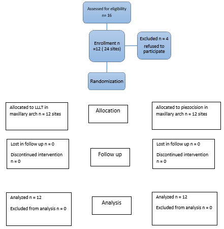
This was a single-center, interventional, double-blind study that was approved by the Institutional Ethical Committee with approval no. IEC/1/2023 and was conducted from Feb 2023 to September 2023 at a government multispecialty hospital using a randomised clinical trial design with a split-mouth technique[R1] [AJ2] and the trial was registered in the CTRI with Ref no. REF/2023/04/082869. Since both the experimental groups were formed from the same patient sample so biological variation[R3] was almost negligible. The research plan is displayed in [Figure 1].[R4]
The distribution of the subjects with respect to age and sex distribution is shown in (false)
Table 1
The treatment provider and participants were blinded till the day ofinterventions, as the chits were not shown to them.
Table 2
The distribution of the subjects with respect to age and sex distribution.
|
Age |
Male |
Female |
|
18 |
0 |
2 |
|
21 |
0 |
1 |
|
22 |
0 |
1 |
|
24 |
1 |
3 |
|
25 |
0 |
1 |
|
26 |
1 |
2 |
|
Total |
2 |
10 |
The patients included in this study belonged to the age group of 15 to 30 years with Class II Div 1 malocclusion requiring first premolar extraction and bilateral maxillary canine retraction. They were also required to have good oral hygiene with gingival sulcus depth not exceeding 3mm which was measured using William’s probe and no previous history of orthodontic treatment.
The exclusion criteria included history of recent periodontal therapy, Congenital disorders, syndromes, cleft lip and palate, patients on systemic corticosteroids and/or anti-inflammatory drugs and Systemic diseases – congenital heart disease, diabetes mellitus, osteoporosis, hyper parathyroidism, Vit D deficiency
Randomization and Blinding
16 patients were assessed out of which 12 met the inclusion criteria. Randomization of side selection for piezocision and LLLT was done by chit method with an allocation ratio of 1:1. Two opaque folded chits were presented to the patient with intervention and side written inside and one was picked up by the patient. A trained dentist carried out randomization and allocation who himself was not involved in treatment part. The allocation of intervention is explained in (Table 2). as the chits were not shown to them.
Interventions
All pre-treatment patient records including lateral cephalogram, OPG (NewTom GIANO, Italy) was used with settings( OPG- 70-80 kV ,8-15 mA & 20 sec and lat. Ceph- 85kV,10mA and 0.5-1 sec).Pre- treatment p0.022 x 0.028-inchlevellinglevellingRound wires were used for retraction as they provide less friction.
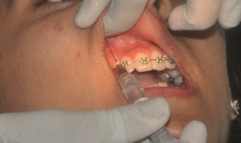
Topical anaesthesia followed by local infiltration (Septodont, France) with 2 % Lignocaine HCL with adrenaline 1:100000 was used before performing intervention as shown in (Figure 2)
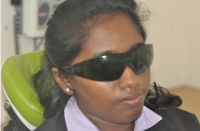
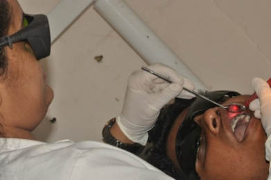
Following procedure was used for the Laser side: A diode laser emitting infrared radiation at 975 nm and functioning in a continuous wave mode was usedstopwatchteeth.8
This was done in a sweeping manner. The tooth's apical third received an 8-second radiation, while the cervical and middle thirds had a 10-second exposure.quadrants.8
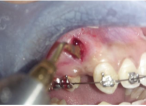
Following procedure was used for piezocision:Piezocision was performed by making a single vertical incision distal to canine, starting 3mm apical to marginal gingiva to preserve gingival papilla and extending 10 mm apically, using a 15 no blade with BP blade handle. Piezotome (Dmetec, Korea) was used to make cortical cuts of 3mm depth in entire length of incision as shown in (Figure 5)
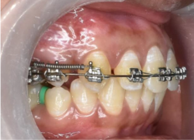
Chlorhexidine 0.12% oral rinse.Maxillary canine retraction was then initiated after 10 days of surgical procedures on 0.018 inch round Australian SS archwire using a NiTi closing coil spring (Leone, Italy) extending from hook on maxillary first molar to the power arm of maxillary canine applying a force of approximately 150gm calibrated with a Dauntrix gauge as shown in (Figure 6). Rate of canine retraction was followed up till 16, 12 weeks and 16r 4 weeks (T1), 8 weeks (T2),
Measurement of tooth movement
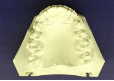
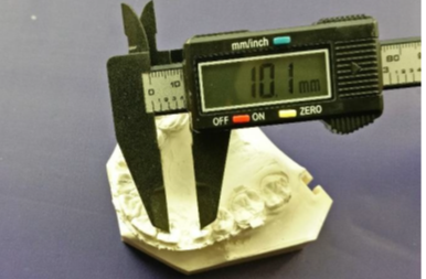
Vertical lines were marked on the casts of each follow up time interval in the middle of the palatal surfaces of canine and lateral incisor, from incisal edge/cusp tip [R1] of the tooth extending to the cervical region. Then the canine retraction was measured at the middle third of the crown using a digital calliper (Mitutoyo, Japan). Figure 7, Figure 8).
To check intra-observer reliability, measurements from 30 randomly selected study models were recorded again after a period of 15 days. The measurements were compiled in excel sheet and statistical analysis was performed with t test and [R4] repeated measures ANOVA [R5] [AJ6] using SPSS software (version 18).
Results
Among the 12 participants there were 2 males (16.6%) and 10 females (83.3%).
Table 3
The comparison of rate of canine retraction in both the experimental groupsare shown in (Table 3)
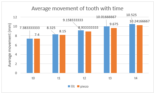
The LLLT group showed 1.105 times the canine retraction in 4 months as compared to the group subjected to piezocision and the findings are statistically significant (P Value = 0.09).Moreover, the mean rate of distal movement was 0.84mm/month and 0.75mm/month in LLLT and piezocision group respectively.
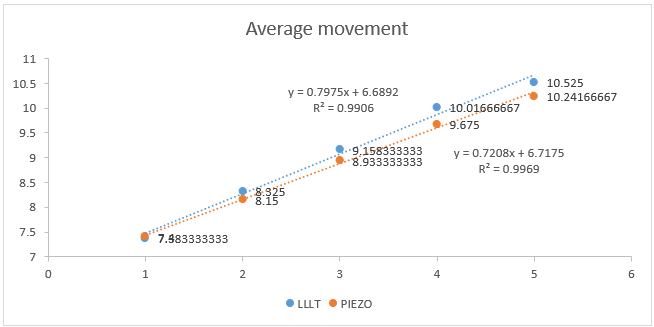
From results we can also infer that, there is a constant decline in the rate of tooth movement throughout the duration of the study, where the rate of canine retraction in the last month is almost half of that seen in the first month of the study as seen in (Figure 9)
The rate of canine retraction is consistently more in the experimental group subjected to LLLT as compared to the group subjected to piezocision in the duration of the study as seen in (Table 4 and Figure 10)
Table 4
The rate of canine retraction is consistently more in the experimentalgroup subjected to LLLT as compared to the group subjected to piezocision inthe duration of the study as seen in (Table 4 and Figure 10).
Discussion
Various surgical techniques are now available in contemporary orthodontic practice that can be used for accelerating the orthodontic tooth movement. Among these techniques Piezocision and LLLT have been considered minimally invasive, easy to perform and better tolerated by the patients.7, 9, 10, 11 In the present study both these procedures were directly compared in terms of rate of canine retraction by using a split mouth study design.
Split mouth study design in general has several advantages including reduction of sample size to half and less risk of bias as described by Pandis.12 Sample size is decreased as each participant provides two interventional sites and since the same participants are providing experimental as well as the control sites the variables associated with biologic factors, age and gender differences, compliance etc. are greatly reduced. However, a patient cannot be enrolled if the two intra oral sides are not similar and such a design is not suitable for evaluating interventions that are not restricted to localized sites and have a generalized effect on both the sides. The above mentioned features of a split mouth study design made it suitable for our study. It provided a direct comparison between the experimental groups by removing the patient related biologic confounders.
The patient selection process was limited to a sample of people between the ages of 15 and 30 years in order to ensure a homogenous sample. It has been discovered that the age of the patient can influence how quickly tooth movement occurs, with younger individuals (less than 15 years old) showing faster tooth movement than older patients. To eliminate the impact of this confounding factor, we limited the number of patients in our trial to those who were older than 15 years.13
Another complicating factor that may impact the rate of cementum and bone remodeling, as well as tooth movement, is the amount of sex hormones in women during the estrous cycle.14, 15 Unfortunately, due to the small number of subjects who agreed to take part in this study, this variable could not be eliminated.
In the present study extraction of first premolars was done before starting treatment followed by leveling and alignment stage which took 12 to 16 weeks on an average. This was done to reduce the bias that may arise from RAP generated because of extractions. This was in accordance with Alfawal et al. 5, Feizbakhsh et al. 16 and Attri et al. 10 The RAP generated was assumed to accelerate tooth movement during leveling and alignment stage and by the time canine retraction was started it would have reduced significantly or completely subsided.
Until now, most investigations have used a lower wavelength spectrum laser operating in the 780-980 nm range, with 810 nm being the most often used wavelength. 17, 18, 19 The first study to investigate the impact of a 980 nm laser on orthodontic tooth movement was conducted in 2016 by Yassaei S et al. 20 Using the same parameter, a previously unpublished study carried out by the authors, demonstrated that applying LLLT with 975 nm laser resulted in 52% faster tooth movement compared to conventional orthodontics. Thus, in this study also 975 nm wavelength was used.
The laser's effect is also influenced by the way the irradiation is delivered. Although the use of the continuous mode has been supported by Bradley et al. (2000) and Takeda et al. (1988), Kim et al. (2009) and Ng D et al. (2017) have recommended the use of the pulsed mode. According to Yoshida T. et al., laser units operating continuously have a higher level of biostimulatory response. 18, 19, 20, 21 Consequently, a continuous mode of operation was used for the irradiations in this investigation. Since cells are more easily influenced by LLLT in the early stages of biological response, Ozawa Y et al., Saito S, and Shimizu N suggested using laser more frequently in the initial phase of tooth movement. 22, 23 Furthermore, it has been proposed by Khadra M et al. and Ng D et al. that multiple doses are preferable to a single dose. 18, 24 In this investigation, laser irradiation was carried out on days 0, 7, and 14 of the first month and then every 15 days for the next three months.
Piezocision was introduced for the first time by Dibart et al5 as a conservative surgical approach for accelerated orthodontics. The procedure involved local anesthesia administration, soft tissue incisions followed by making about 3mm deep buccal cortical cuts using a piezotome through these incisions without elevating a flap.
In our study also Piezocision (Figure 5, Figure 6) was performed similar to the procedure described above. The soft tissue incision was made starting 3mm away from marginal gingiva. The sparing of marginal gingiva and associated bone from surgical trauma was done to prevent alveolar bone loss and gingival recession as has been described by Abbas et al.3, Sebaoun et al.25 and Hoogeveen et al.26 The piezocision cuts extended 10mm in length distal to canine
In our study the experimental group subjected to LLLT showed 1.105 times the canine retraction in 4 months as compared to the group subjected to piezocision.
Angiogenesis brought on by LLLT exposure promotes the quick removal of agents that cause resorption. 17, 18 Additionally, LLLT speeds up the rate of remodelling whereby anabolic activity outpaces catabolic activity.
Piezocision works on the mechanism of regional acceleratory phenomenon (RAP). The acceleration of orthodontic tooth movement is explained by increased activity of underlying molecular mechanism. A number of studies have demonstrated the role of inflammatory markers like chemokines and cytokines in OTM and the fact that their levels are significantly increased following traumatic insult to the periodontium.27, 28, 29, 30, 31 These raised levels of inflammatory markers can be maintained further by application of orthodontic forces. Chemokines are involved in the recruitment process of osteoclast precursor cells and cytokines induce maturation of osteoclasts from their precursor cells through prostaglandin E2 and RANK/RANKL/OPG pathway. Therefore, it has been established that increasing the expression of these factors by surgically insulting the bone accelerates tooth movement.
From our study we can also discern that the rate of canine retraction was maximum in the 1st month and least in the last month. There is a constant decline in the rate of tooth movement throughout the duration of the study, where the rate of canine retraction in the last month is almost half of that seen in the first month of the study. The canine showed a mean distal movement of 3.14mm and 2.84 mm in LLLT and piezocision group respectively.
Conclusion
LLLT and piezocision are widely accepted minimally invasive acceleratory orthodontic procedures. The rate of tooth movement is slightly higher in teeth exposed to LLLT. With no need of incision and lesser chance of damage to hard and soft tissue, LLLT appears to be a comparatively attractive option for acceleratory orthodontics. However further studies with larger sample sizes are needed to ascertain possible side effects using the split-mouth technique. Studies are also needed to evaluate the effects of acceleratory orthodontics in populations staying in challenging topography like high altitude areas, where the local environmental factors dampen the tooth movement.

