- Visibility 587 Views
- Downloads 53 Downloads
- Permissions
- DOI 10.18231/j.jco.2022.028
-
CrossMark

Abstract
Atraumatic extractions are always the prerequisite for immaculate orthodontic results. Of all the extractions desired by the orthodontist, maxillary first premolar is challenging for the dentist / oral surgeon specifically because of its thin and slender roots with wide variations in its anatomy. Various methods for atraumatic premolar extraction and for preserving the buccal cortical plate have been suggested in the past like use of periotomes, physics forceps etc. Following is the suggested novel technique for atraumatic premolar extractions which can be performed with routine forceps without any additional armamentarium.
Introduction
Elective extractions of premolars are routinely performed for facilitating orthodontic treatment. Among these, the maxillary first premolars have extremely thin roots and tend to fracture, especially in adult patients with high bone mineral density and decreased bone elasticity. Perhaps the most common root fracture when extracting teeth in adults occurs with this tooth.[1]
Unforeseen complications while extraction of maxillary first premolars has been widely discussed in the literature. The three most common intraoperative complications reported in the literature are crown fracture, root fracture, and fracture of alveolar cortices due to numerous differences in the surrounding bone type[2] and thickness. The reported frequency of root fracture ranged from 5% to 7% for all extractions but was as high as 30% in cases of dilacerated and divergent roots of maxillary premolars.[3]
Importance of atraumatic extraction while preserving the buccal cortical plates in orthodontic extraction cannot be tossed aside. Different techniques for atraumatic extractions are suggested in literature, such as use of periotomes,[4] piezosurgery,[5] physics forceps,[6] etc. all these methods need expertise, special instrumentation, have limited applications and may result in iatrogenic effects such as causing laceration to gingiva and buccal mucosa, ulcer formation, etc.[7]
An effective clinical technique, which eases tooth removal while preserving the surrounding hard and soft tissues, is suggested there forth.
Procedure
The technique is demonstrated in a 22 years old female patient who required first premolar extractions for her orthodontic treatment. Pre- treatment intraoral photograph of the patient was taken. ([Figure 1]) Patient was bonded with MBT prescription .022slot with the usual/ required protocol except for the maxillary first premolars which were bonded 0.5 -1mm gingival to ideal vertical position. ([Figure 2] ) Occlusal reduction of 0.5- 1 mm was performed on the first premolars with round end tapered diamond burs.[8] ([Figure 2], [Figure 3] ) The initial wire placed was 0.014”


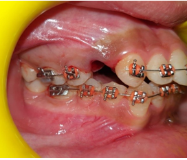
HANT (heat activated nickel titanium) for light continuous force. ([Figure 4] ) Patient was recalled after 2 weeks. Arch wire was sectioned distal to canine and mesial to second premolar. First premolar bracket was deboned. ([Figure 5] ) Then the maxillary First pre molar Extractions were performed using routine forceps with simple extrusive and mild jiggling moments. ([Figure 6])
Discussion
Orthodontic extractions are conventionally done preceding the formal bonding procedure. However, it’s suggested that cases intended for extraction, specifically in some selected adult patients or cases where difficult extractions are anticipated, extraction may be postponed for two weeks or more (to facilitate above technique), as the clinical situations warrants.
The placement of bracket gingivally by 0.5 to 1 mm on the premolar and disocclusion of the premolars by occlusal reduction helps in their free movement, in all three planes of space. Force applied with initial wire leads to a slight widening of periodontal space which is clinically observed as a mild loosening of the tooth in its socket. The extractions thus performed are atraumatic, preserving the buccal cortical plates which are prerequisite for impeccable orthodontics results.
Source of Funding
None.
Conflict of Interest
None.
References
- Ash MM. Wheeler's Dental anatomy, physiology, and occlusion. . 1993. [Google Scholar]
- Misch CE. Contemporary implant dentistry. . 1993. [Google Scholar]
- Narendar R, Balakrishnan G, Kavin T, Venkataraman S, Altaf S, Gokulanathan S. Incidence of Risk and Complications Associated with Orthodontic Therapeutic Extraction. J Pharm Bioallied Sci. 2017;9(1):201-4. [Google Scholar]
- Levitt D. Atraumatic extraction and root retrieval using the periotome: a precursor to immediate placement of dental implants. Dent Today. Future Dent J. 2001;20(1). [Google Scholar]
- Stübinger S, Kuttenberger J, Filippi A, Sader R, Zeilhofer H. Intraoral piezosurgery: preliminary results of a new technique. J Oral Maxillofac Surg. 2005;63(9):1283-7. [Google Scholar]
- Golden RM. Dental plier design with offsetting jaw and pad elements for assisting in removing upper and lower teeth utilizing the dental plier design. US patent 6,910,89. . 2005. [Google Scholar]
- Kapila S, Kaur T, Bhullar R, Sandhu A, Dhawan A, Kaur A. Use of Physics Forceps in Atraumatic Orthodontic Extractions of Bilateral Premolars: A Randomized Control Clinical Study. J Maxillofac Oral Surg. 2020;19(3):347-54. [Google Scholar]
- Shillingburg H. Fundamentals of fixed prosthodontics. . 1997. [Google Scholar]