- Visibility 431 Views
- Downloads 77 Downloads
- DOI 10.18231/j.jco.2022.030
-
CrossMark
- Citation
Correlation between cephalic index and facial index in skeletal malocclusions – An analytical cross-sectional study
- Author Details:
-
Prachodh C A
-
Vincy Antony *
-
Muhamed Shaloob
-
Prathapan Parayaruthottam
-
Mohamed Nayaz
-
Muhammed Raheesh
Introduction
The assessment of facial form and cranial morphology are very important in planning orthodontic treatment and its prognosis. Retzius G[1] in 1840 gave the first classification based on cranial morphology. When used in living individuals, these craniofacial measures are referred to as Cephalic Index, whereas Cranial Index is used when referring to dry skulls. Ricketts[2] in 1964 introduced the terms dolichofacial, brachyfacial and mesofacial. According to Ricketts,[2] mesofacial describes patients with Class I malocclusion having a pleasant soft tissue profile, an average facial pattern and a normal maxillo-mandibular relationship. A horizontal growth pattern usually associated with Class II Division 2 malocclusion is referred to as brachyfacial type. Dolichofacial type which is associated with Class II Division 1 malocclusion, usually presents with a vertical growth pattern. The term Facial Index is used to represent facial proportions[3] The Facial Index was determined by dividing the Nasion-Menton length by the interzygomatic width.
The null hypothesis stated that there was no correlation between cephalic and facial indices in the different skeletal antero-posterior malocclusions. The following article investigated the possible influence of the cranial morphology on the facial type in patients with skeletal malocclusions.
Materials and Methods
This analytical cross-sectional study was approved by the Institutional Ethical Committee (IEC/MES/66/2019 dated 20/11/19) and followed the criteria in the 2013 Declaration of Helsinki. The study sample was selected after fulfilling the requirements for inclusion and exclusion criteria. A written informed consent was obtained before the photographs of the patients were taken as a part of the study.
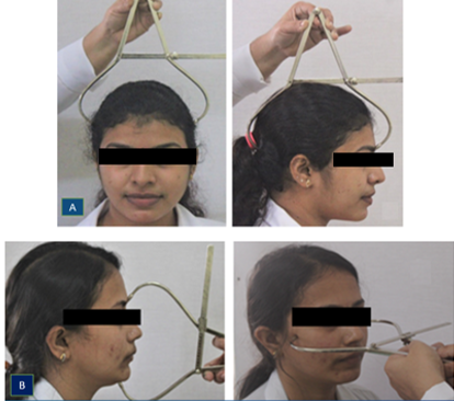
Patients between ages 18 – 30 years, with intact molar and canine relationship in the maxillary and mandibular dentition and having different antero-posterior skeletal malocclusions, and their corresponding dental malocclusion were included in the study ([Figure 1]). Patients with any cranial and dental anomalies, prior orthodontic therapy, history of trauma in the cephalic and facial region were excluded from the study.


|
Head type |
Cephalic Index |
Calculation |
|
Hyperdolichocephalic |
65.0-69.9 |
The Cephalic Index (CI) was calculated with the formula:[4] |
|
Dolichocephalic |
70.0 – 74.9 |
|
|
Mesocephalic |
75.0 – 79.9 |
CI = Maximum head width (Eu') – (Eu')x 100/ Maximum head length (G -Op) |
|
Brachycephalic |
80.0 – 84.9 |
|
|
Hyperbrachycephalic |
85.0 – 89.9 |
|
|
Ultrabrachycephalic |
≥ 90.0 |
|
|
Face type |
Facial index |
Calculation |
|
Hypereuryprosopic |
≤79.9 |
The Facial Index (FI) was calculated by the formula:[4] |
|
Euryprosopic |
80.0 – 84.9 |
|
|
Mesoprosopic |
85.0 – 89.9 |
FI = Facial height N -Me × 100Bizygomatic face width |
|
Leptoprosopic |
90.0 – 94.9 |
|
|
Hyperleptoprosopic |
≥ 95.0 |
|
Parameter |
Class I |
Class II |
Class III |
|||
|
(%) |
|
(%) |
|
(%) |
|
|
|
Cephalic index |
Number |
Number |
Number |
|||
|
Hyperdolichocephalic |
7.5 |
3 |
2.5 |
1 |
5 |
2 |
|
Dolichocephalic |
12.5 |
5 |
5 |
2 |
32.5 |
13 |
|
Mesocephalic |
55 |
22 |
57.5 |
23 |
42.5 |
17 |
|
Brachycephalic |
20 |
8 |
32.5 |
13 |
17.5 |
7 |
|
Ultrabrachycephalic |
5 |
2 |
2.5 |
1 |
2.5 |
1 |
|
Facial Index |
||||||
|
Hypereuryprosopic |
0 |
0 |
0 |
0 |
0 |
0 |
|
Euryprosopic |
7.5 |
3 |
5 |
2 |
7.5 |
3 |
|
Mesoprosopic |
50 |
20 |
57 |
23 |
7.5 |
3 |
|
Leptoprosopic |
22.5 |
9 |
27.5 |
11 |
27.5 |
11 |
|
Hyperleptoprosopic |
20 |
8 |
10 |
4 |
57.5 |
23 |
|
Groups |
Class I |
Class II |
Class III |
|
Cephalic Index |
80.77 ± 5.91 |
79 ± 3.89 |
77.84 ± 5.57 |
|
Facial Index |
87.17 ± 5.57 |
87.04 ± 5.27 |
93.08 ± 4.26 |
|
T Value |
5.01 |
7.85 |
13.92 |
|
P value |
0.0001* |
0.0001* |
0.0001* |
|
Groups |
Class 1 |
Class II |
Class III |
|
Cephalic Index |
-0.28(P=0.08) |
-0.15(P=0.35) |
-0.08(P=0.62) |
|
Facial Index |
Study sample
The sample size was calculated in the present study using a two-sided test to detect correlation r (Estimated correlation coefficient r= 0.637), α= 0.05, β= 0.1, the calculated sample size was n > 22; the sample size taken was 40 in each group.
120 patients were selected for the study who were split into three groups, each group containing 40 patients. Each group had patients with their corresponding sagittal skeletal and dental malocclusion. All the participants were examined in the dental chair with head kept oriented to the Frankfort-horizontal plane. The patients were classified into different sagittal malocclusions based on Clinical and Cephalometric assessment as shown in [Figure 5].
Evaluation of Cephalic and Facial Index
Cephalic index: The Cephalic Index refers to a ratio between the width of head and length of head.[4] In the present study, a standard spreading caliper was used for taking the measurements of head width and length ([Figure 1]) for the estimation of Cephalic Index. The Cephalic Index was measured using the landmarks[4] Eurion (Eu′), Opisthocranion (Op), Glabella (G′).
Facial index: The ratio between the facial height to the bizygomatic facial width describes the facial index.[4] In this study, a standard spreading caliper ([Figure 1]) was used to measure facial height and facial width. The different landmarks used for the measurement of the Facial Index were Nasion (N′), Menton (Me′), Zygion (Zy′). The participants in this study were categorized according to Martin and Saller’s classification for Cephalic and Facial index ([Table 1] ). All linear measurements were recorded in millimeters to 0.10” accuracy. To control any measurement error, all measurements were taken twice and if there was a discrepancy, a third reading was taken.
Results
The study consisted of 120 participants, with 57 male patients and 63 female participants. Males contributed to 47.5% and females contributed to 52.5% of the total study population. The statistical analyses were done by using the SPSS (Version 22-SPSS Statistics for Windows, IBM Corp., Armonk, NY, USA). The level of significance was set at p<0.05. The Shapiro-Wilk test was used to test the normality of the data. The independent t-test was used to calculate any difference between the 2 groups and Chi-square test was used for proportion analysis. Correlation coefficient between the Facial index and Cephalic index was calculated using Pearson Correlation Coefficient. The confidence interval of 95%, the Power of the study at 80%, and the probability of α-Error at 5 % were selected.
While analysing the Cephalic indices of the study subjects in different antero-posterior malocclusions ([Table 2]), it was found that Mesocephalic head form was the predominant head form in Class I (55%), Class II (57.5%) and Class III (42.5%) malocclusions. The least prevalent was Ultrabrachycephalic - 5% in Class I and 2.5% in both Class II and Class III malocclusions respectively. While evaluating Facial indices for the different antero-posterior malocclusions ([Table 2]), it was found that Mesoprosopic was most predominant in Class I and Class II with an incidence of 50 % in Class I, 57 % in Class II, and 7.5 % in Class III malocclusion.
After analyses and comparison of the mean values for Cephalic Index and Facial Index using independent t-test, a difference which was statistically significant was observed between the two indices in all the study groups (p<0.05) as seen in [Table 3].
Discussion
The present study employed anthropometric/craniofacial measures which are widely used to describe and classify the face and head form according to Martin and Saller. Cephalic index and Facial index of 120 adult patients were evaluated.
Head form prevalence: The results indicated a prevalence of Mesocephalic head form (45.1%) in the Malabar region of Kerala. Concordant results with predominance of mesocephalic head form were observed in the studies of Njemirovskij V et al.,[5] Alves HA et al,[6] Nair SK et al,[7] Patro S et al,[8] Mishra M et al,[9] Akinbami BO,[10] Lakshmi KK et al,[11] Shah T et al,[12] Setiya M et al,[13] Ahmed SKN and Sreenivasan M,[14] Ranga MKS and Mallika MCV,[15] Doshi MA and Jadhav SD,[16] Mangeshkar A et al.[17] and Thomas MW and Rajan SK.[18] The present study results showed Brachycephalic head form as the second most prevalent group with an incidence of 21% among the study subjects. Dolichocephalic head form was the third most (7.1%) common in the study population. The prevalence of the Hyperdolichocephalic type was 4%. The rarest head type in this study was found to be the Ultrabrachycephalic head type (3%).
Face form prevalence: While evaluating the Facial index, the Mesoprosopic face form was the prevalent group with an incidence of 38.3%, thus indicating a predominance of Mesoprosopic face form in the Malabar region of Kerala. The predominance of Mesoprosopic face form was also seen in the studies done by Njemirovskij V et al,[5] Kumar M and Lone MM,[19] Prasanna PL et al.[20] Hyperleptoprosopic face form was the second most prevalent in our study population with an incidence of 31.8%. Unlike our study, the Hyperleptoprosopic face form were found to be the most predominant face type as reported by Kamble NB and Kamble D,[21] Maina MB et al.[22] and Kataria DS et al.[23] Leptoprosopic face form was the third most prevalent in the study population with an incidence of 25.8%. In the present study incidence of Euryprosopic face form was 10.8%.
Cephalic index in different antero-posterior malocclusions: While evaluating the prevalence of Cephalic index in different skeletal malocclusions, it was observed that the Mesocephalic head form predominated in all three sagittal malocclusions, with an incidence of 55% in Class I, 57.5% in Class II, 42.5% in Class III malocclusion. Similar to the present study, Rao NR et al.[24] also observed a predominance of Mesocephalic head form in Class I subjects in their study. The current study gave an incidence of Brachycephalic head form of 20% in Class I, 32.5% in Class II, 17.5 % in Class III malocclusion. Dolichocephalic -12.5 % in Class I, 5% in Class II, 32.5 % in Class III malocclusion. Hyperdolichocephalic head form showed an incidence of 7.5% in Class I, 2.5% in Class II, 5% in Class III malocclusion. The least prevalent was Ultrabrachycephalic - 5% in Class I, 2.5% in Class II, 2.5% in Class III malocclusion. Rao NR et al.[24] had observed that Brachycephalic head were more prevalent in skeletal and dental Class III or Class I occlusion patterns and Dolichocephalic head corresponded with skeletal and dental Class II occlusion pattern.
Facial index in different antero-posterior malocclusions: Mesoprosopic face form predominated in Class I and Class II malocclusion with an incidence of 50 % in Class I, 57% in Class II, and 7.5 % in Class III malocclusion. Class III malocclusion showed a higher prevalence of Hyperleptoprosopic face (57.5%) than Class II (10%) and Class I (20%) malocclusion. The incidence of Leptoprosopic was 22.5 % in Class I, 27.5 % in Class II and Class III malocclusion. The least prevalent was Euryprosopic with an incidence of 7.5% in Class I, 5% in Class II, 7.5% in Class III malocclusion. The study done by Rao NR et al.[24] observed that Mesoprosopic face was more associated with skeletal and dental Class I pattern which was in concordance with our findings however, they also reported that Leptoprosopic face showed more skeletal and dental Class II occlusion pattern and that Euryprosopic face had skeletal and dental Class III or Class I occlusion pattern which was not evident in our study.
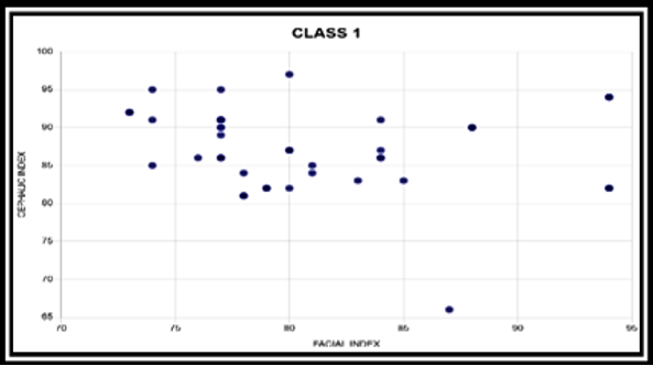
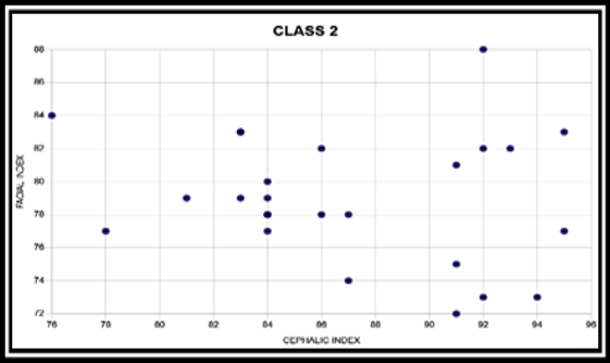
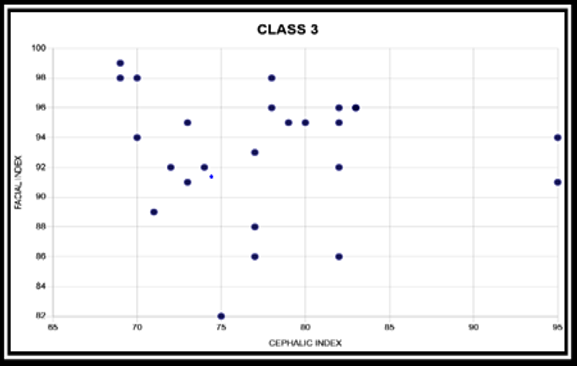
Correlation between head form and face form: The strength of the linear relationship between two variables is quantified as the correlation coefficient. The concordance between Facial Index and Cephalic Index was analysed to observe the correlation between them if present. Results of the present study demonstrated a weak negative correlation between the Cephalic Index and Facial Index in different antero-posterior malocclusions - (-0.28) in Class I malocclusion, (-0.15) in Class II malocclusion and (-0.08) in Class III malocclusion ([Table 4] and [Figure 2], [Figure 3], [Figure 4]). Catharino F et al. Observed that of the study subjects who were classified as brachycephalic, 52.6% were leptoprosopic, whereas only 10.5% were euryprosopic. Catharino F et al.[25] had observed that of the study subjects who were classified as brachycephalic, 52.6% were leptoprosopic, whereas only 10.5% were euryprosopic. Menapace et al.[26] in their study also observed weak association between face form and head form and found frequent association between the euryprosopic facial type and the dolichocephalic head shape. Raghavendra et al.[27] in 2021 also observed no significant correlation between the cranial and facial parametersin the study subjects, which is in consensus with our study results.
However, there have been studies in the previous literature which support the consensus between cranial and facial morphology. That would support the paradigm that the head and face type would be similar; that is, individuals with a leptoprosopic face form would have a corresponding dolichocephalic head type. According to Bhat M and Enlow DH[28] in 1985, the cranial base serves as a model for the face. Rao NR et al.[24] also observed a positive correlation between the Cephalic and Facial indices.
The present research was limited by a few factors like the sample size. The selection of larger number and more representative sample with different skeletal malocclusion would provide more reliable results. The results are in concordance with our null hypothesis that there is no correlation between facial and cephalic indices in the different antero-posterior malocclusions.
Conclusion
The present study investigated the correlation between head and face forms in patients with different skeletal antero-posterior malocclusions. The study concluded that:
There was a predominance of Mesocephalic head form in the different antero-posterior malocclusions (Class I, Class II and Class III malocclusion).
Among the face form, Mesoprosopic predominated in Class I and Class II malocclusion, Hyperleptoprosopic face was the most common in Class III malocclusion.
A weak negative correlation existed between head form and face form in Class I, Class II and Class III malocclusion. The results indicate that cranial morphology and facial morphology exerts a weak influence on each other.
The study results possibly suggest the prevalence of Mesocephalic head form and Mesoprosopic and Hyperleptoprosopic face form in the Malappuram district of Kerala.
Financial Support and Sponsorship
None.
Conflicts of Interest
There are no conflicts of interest
References
- G Retzius. A Qualitative Analysis of How Anthropologists Interpret the Race Construct. Am Anthropol 1909. [Google Scholar]
- RM Ricketts. The keystone triad:II. Growth, treatment and clinical significance. Am J Orthod 1964. [Google Scholar]
- J Cameron. A study of the upper facial index in diverse racial types of mankind. Craniometric studies. Am J Phys Anthropol 1929. [Google Scholar]
- FB Naini. Facial aesthetics - Concepts and clinical guidelines. 2011. [Google Scholar]
- V Njemirovskij, Z Radović, D Komar, B Lazić, T Kuna. Distribution of craniofacial variables in south Dalmatian and middle Croatian populations. Coll Antropol 2000. [Google Scholar]
- HA Alves, M Santos, F Melo, R Wellington. Comparative study of the cephalic index of the population from the regions of the North and South of Brazil. Int J Morphol 2011. [Google Scholar]
- SK Nair, VP Anjankar, S Singh, M Bindra, D Satpathy. The study of cephalic index of medical students of central India. Asian J Biomed Pharm Sci 2014. [Google Scholar]
- S Patro, R Sahu, S Rath. Study of cephalic index in Southern Odisha population. J Dent Med Sci 2014. [Google Scholar]
- M Mishra, A Tiwari, DC Naik. Study of cephalic index in Vindhya region of Madhya Pradesh. Int J Med Sci Public Health 2014. [Google Scholar]
- BO Akinbami. Measurement of cephalic indices in older children and adolescents of a Nigerian population. Bio Med Res Int 2014. [Google Scholar]
- KL Kumari, PV Babu, PK Kumari, M Nagamani. A study of cephalic index and facial index in Visakhapatnam. Int J Res Med Sci 2015. [Google Scholar]
- T Shah, MB Thaker, SK Menon. Assessment of cephalic and facial indices: a proof for ethnic and sexual dimorphism. J Forensic Sci Criminol 2015. [Google Scholar]
- M Setiya, A Tiwari, M Jehan. Morphometric Estimation of Cranial Index in Mahakoushal Region of Madhya Pradesh: Craniometrics Study. Int J Sci Stud 2018. [Google Scholar]
- SK Ahmed, M Sreenivasan. Study of Cephalic Index among the Tamil Population. Medico Legal Update 2019. [Google Scholar]
- MKS Ranga, MCV Mallika. Cephalic index and facial index of adults in rural South Kerala, India. Int J Sci Stud 2020. [Google Scholar]
- MA Doshi, SD Jadhav. Cephalic Index And Head Shape In Western Maharashtra Students. Res J Pharm Biol Chem Sci 2020. [Google Scholar]
- A Mangeshkar, AB Najan, VK Gohiya, S Gohiya. Estimation of Cephalic index in 17-20 Years old population of Nimad region of Madhya Pradesh. Indian J Forensic Med Toxicol 2021. [Google Scholar]
- MW Thomas, SK Rajan. Regional and Gender Differences in the Cephalic Index among South Indian and North-East Indian Population. Medico Legal Update 2021. [Google Scholar]
- M Kumar, MM Lone. The Study of Facial Index among Haryanvi Adults. Int J Sci Res 2013. [Google Scholar]
- PL Prasanna, P Suresh, K Srinivasan. Anthropometric Study of the Facial (Prosopic) Indices: A Proof for Gender Dimorphism. J Dent Educ 2020. [Google Scholar]
- NB Kamble, D Kamble. Anthropometric Study of Cephalic and Facial Indices among Central Indian Population. Indian Internet J Forensic Med Toxicol 2020. [Google Scholar]
- MB Maina, O Mahdi, GG Kalayi. Craniofacial forms among three dominant ethnic groups of Gombe State, Nigeria. Int J Morphol 2012. [Google Scholar]
- DS Kataria, RK Ranjan, SA Perwaiz. Study of variation in total facial index of north Indian population. Int J Health Sci Res 2015. [Google Scholar]
- NR Rao, CB More, S Patel. Correlation of cephalic index, facial index with skeletal and dental malocclusion using morphologically measurable parameters in males and females. Int J Curr Res 2016. [Google Scholar]
- F Catharino, D Feu, A Rossi, M Normando, D Martins De Araujo. Cranial morphology and facial type: Is it appropriate to describe the face using skull terminology?. J World Fed Orthod 2014. [Google Scholar]
- SE Menapace, DJ Rinchuse, T Zullo, CJ Pierce, H Shnorhokian. The dentofacial morphology of bruxers versus non-bruxers. Angle Orthod 1994. [Google Scholar]
- AY Raghavendra, SM Bhosale, SK Naik, MA Janvekar. Determination of Craniofacial Relation in Human Dry Skulls: An Anthropometric Study. Int J Health Clin Res 2021. [Google Scholar]
- M Bhat, DH Enlow. Facial variations related to head form type. Angle Orthod 1985. [Google Scholar]
