- Visibility 172 Views
- Downloads 8 Downloads
- DOI 10.18231/j.jco.2023.026
-
CrossMark

Management of deepbite in Class II division 2 malocclusion with two different techniques- A case series
- Author Details:
-
 Mukesh Kumar
-
Manish Goyal
-
Haripriya Nongthombam *
-
Sumit Kumar
Introduction
The treatment of Class II division 2 malocclusion is usually known to be challenging and prone to relapse.[1] The cases of this type of malocclusion are usually characterized by severe, traumatic deep overbite with palatally inclined maxillary incisors. Growth modification has long been used to treat Class II malocclusion in teenagers. The treatment option, such as functional appliances, aim to restrict or divert maxillary growth while also increasing mandibular growth.[2], [3] On the other hand, orthognathic surgery is typically the only option for non-growing patients with severe Class II malocclusions, which usually have severely retrognathic mandible.[4] In conditions where the above-mentioned treatment options are infeasible, orthodontic camouflage with correction of the deep bite by intrusion of incisors, extrusion of the molars or both, and proclination of the incisors can be considered.
The treatment therapy objectives, according to Uribe and Nanda,[4] should include the patient's major concern, and the mechanics of correction should be personalized to each patient's needs and goals. In non-extraction cases, resolution of the discrepancy is generally achieved through lateral expansion of the dentition, labial movement of the incisors, and molar distalization. However, the use of these techniques is limited by the patient’s maxillofacial morphology and stability.
Orthodontic camouflage, with extraction of premolars, is another treatment option usually considered. Extraction of all first premolars / extraction of maxillary first premolars and mandibular second premolars / extraction of maxillary second molars for maxillary arch distalization / extraction of maxillary premolars with mandibular incisors, or even extraction of a single mandibular incisor, has all been proposed as extraction patterns. Periodic examination of changes during the treatment is required.[4]
In this article, we have discussed the management of two cases of Class II, division 2 malocclusion patients treated with upper first premolar extraction with different treatment mechanics.
Case 1

Diagnosis
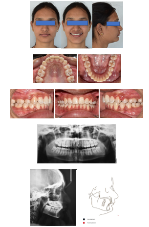
A 14-year-old male presented with the chief complaint of irregularly placed front teeth. Extra oral clinical analysis showed a convex facial profile with no significant facial asymmetry, mesoprosopic facial type, posterior facial divergence, incompetent lips and acute nasolabial angle. Intraorally, there was Class II molar relationship on the left side and end-on molar relation on the right side with 1 mm of overjet and complete deepbite with buccally erupted canines in the upper arch (13, 23). Mild crowding was observed in the lower arch. An amalgam restoration was present in relation to maxillary right permanent first molar (16) and a buccal pit caries was observed with respect to mandibular left permanent first molar (36) ([Figure 1]).
Radiographic evaluation revealed the presence of all four third molars with root formation remaining. Lateral cephalometric analysis revealed a skeletal Class II malocclusion (ANB, 4°), with hyperdivergent jaw bases (SN-Go-Me, 33°), with retroclined upper and proclined lower incisors (U1 to N-A, 21°/1 mm, L1 to N-B, 27°/4 mm).
Treatment objectives
Treatment goals, in this case, were to
Level and align both upper and lower arches,
Correct incisor inclinations,
Achieve a Class II molar relationship on the right while maintaining Class II molar relationship on the left,
Achieve optimum overjet, overbite and soft tissue profile.
Treatment alternative and plan
The treatment plans considered were
Fixed orthodontic treatment with extraction of upper first premolars and end up in Class II molar and Class I canine relationship bilaterally.
Fixed orthodontic treatment with extraction of all first premolars followed by fixed functional appliance and end up in Class I molar and canine relationship bilaterally.
The first treatment option was selected as the final treatment plan.

Treatment progress
The upper first premolars were extracted, and the upper arch was bonded using a pre-adjusted edgewise fixed appliance (0.022′′ x 0.028′′ slot, 3M Unitek) with MBT prescription. For anchorage, a transpalatal arch (0.032′′ stainless steel wire) was put in the maxillary arch. Bonding in the lower arch was postponed until the upper arch had acquired sufficient overjet and alignment.
The upper arch was leveled and aligned with a 0.012′′ nickel titanium archwire (3M Unitek nitinol super elastic, USA), followed by 0.014′′, 0.016′′ nickel titanium archwires (3M Unitek nitinol super elastic, USA). In the upper arch, active canine lacebacks were used bilaterally ([Figure 2] A). An anterior bite plane was added in the upper arch after five months to open the bite and aid in leveling and aligning ([Figure 2] B). It was used for a period of two months. Simultaneously, the lower arch was bonded, and leveling and alignment began with 0.014′′ nickel titanium archwire (3M Unitek nitinol super elastic, USA), gradually moving to wires of higher dimensions.
Most of the extraction space in the upper arch was closed after eight months of leveling and alignment as the ectopically erupted canines aligned in arch, and the transpalatal arch was also removed during this time to facilitate the mesial movement of the left maxillary molar. On 0.016"x0.022" stainless steel (SS American Orthodontics), a continuous intrusion arch of 0.016″ Australian A.J. Wilcock archwire was used to correct the upper anterior teeth inclination ([Figure 2] C).
The continuous intrusion arch was used to address overbite by intruding the upper anterior teeth. Following the intrusion of the upper front teeth, settling elastics (3/16′′, 4.5 oz) were utilised bilaterally on a 0.014" nickel titanium archwire (3M Unitek nitinol super elastic, USA) for two months to achieve appropriate occlusion. Fixed upper (2-2) and lower (3-3) lingual retainers were bonded after debonding.
Treatment result
All the treatment objectives were obtained and the patient was satisfied with the outcome. A bilateral Class II molar relationship with optimum overjet, overbite and soft tissue profile was achieved. ([Figure 3]).

Case 2
Diagnosis
A 17-year-old female patient presented with the chief complaint of irregularly placed front teeth. Extraoral examination revealed a mesoprosopic facial form with convex profile, posterior facial divergence, and competent lips. Intraoral examination revealed end on molar and canine relationship bilaterally with 1mm overjet, and complete deep bite with a highly placed upper right canine (13) and retroclined maxillary central incisors (11,21). Mild crowding was seen in both upper and lower anterior region.
Radiographic evaluation revealed the presence of all four third molars. Lateral cephalometric analysis revealed a skeletal Class II malocclusion (ANB, 4°), hypodivergent jaw bases (SN-Go-Me, 29°), and retroclined upper and lower incisors (U1 to N-A, 14°/-4 mm, L1 to N-B, 19° /1mm) ([Figure 4]).

Treatment objectives
Treatment goals were to
Achieve ideal leveling and alignment of both the arches,
Obtain Class II molar relation and Class I canine relation bilaterally,
Achieve optimum overjet, overbite and soft tissue profile.
Treatment alternative and plan
The treatment options were
Orthodontic camouflage with the extraction of the upper first premolars followed by retraction and intrusion of the anterior teeth to correct the complete deep bite.
Fixed orthodontic treatment with BSSO advancement, without extraction of teeth.
Orthodontic camouflage with upper first premolars extraction was chosen as the treatment of choice, as the patient refused surgery.
Treatment progress
The upper first premolars were extracted, and the upper arch was bonded using a 3M Unitek pre-adjusted edgewise fixed appliance (0.022′′ x 0.028′′ slot). For anchorage, a transpalatal arch (0.032′′ stainless steel wire) was put in the maxillary arch. With 0.012′′ nickel titanium archwire (3M Unitek nitinol super elastic, USA), leveling and alignment were started in the upper arch and progressed to wires of increasing thickness. In the upper arch, active canine lace backs were used bilaterally ([Figure 5] A). After six months of leveling and alignment, the ectopically erupted canine (13) aligned in arch, closing most of the extraction area in the first quadrant.
After achieving sufficient alignment in the upper arch, the lower arch was bonded, and leveling and alignment were started with 0.014′′ nickel titanium archwire (3M Unitek nitinol super elastic, USA). As the archwire proceeded to larger dimensions, mild crowding in the lower arch was handled by interproximal reduction. For the intrusion and retraction of the upper central and lateral incisors, a three-piece intrusion arch (0.021′′×0.025′′ stainless steel anterior section and 0.017′′×0.025′′ TMA wire spring) was used ([Figure 5] B).
Four months later, 0.012′′ nickel titanium archwire (3M Unitek nitinol super elastic, USA) was used for intrusion of the upper canine, while a passive utility arch was used to maintain the position of the upper incisors. After three months, the slight midline deviation was corrected with anterior cross elastics (3/16′′, 4.5 oz) on 0.016′′ stainless steel archwire (SS American Orthodontics) ([Figure 5] C). With 0.014′′ nickel titanium archwire, the occlusion was settled in both arches (3M Unitek nitinol super elastic, USA). After debonding, upper lateral to lateral incisor (2-2) fixed retainers and lower removable retainers were provided.

Treatment result
The upper anterior crowding and complete deep bite were resolved. The patient was satisfied with the treatment outcome. Acceptable overjet and overbite were achieved, along with Class I canine and Class II molar relationship bilaterally ([Figure 6]).
Discussion
The orthodontic treatment for Class II division 2 malocclusion is challenging.[1] Various treatment suggestions could be proposed to the same patient with Class II division 2 malocclusion, depending on each orthodontist’s treatment philosophy. Class II maloclussion, division 1 and division 2, are amenable to non-extraction treatment in the late mixed dentition. In these patients, the maxillary arch can usually be adapted to the mandibular arch with part-time headgear therapy and concomitant mandibular growth, assuming favourable sagittal and vertical divergence and transverse skeletal characteristics. Adults with mild to moderate Class II malocclusions require a different treatment approach. One possibility is to distalize the maxillary posterior teeth with extraoral forces or intraoral removable or fixed appliances; second possible treatment approach is to extract teeth only in the maxillary arch and accept a molar distocclusion. Maxillary second premolars extraction is another option.[5]
Because of the possibility for negative consequences on the facial profile, overbite, and reopening of the extraction areas after treatment, it is recommended that caution be exercised with four first premolar extractions.[6] Also, extraction in the mandibular arch is not suggested because correction of overbite has a high risk of relapse.[7] In an extreme case, orthognathic surgery may be required to achieve an ideal correction. However, often, most patients refuse to undergo orthognathic surgery due to financial or other concerns, even though it might be the best treatment for them. Hence, orthodontic camouflage, with extraction or non-extraction, is often the most considered treatment option. Although premolar extractions may be used to achieve camouflage, the soft-tissue objectives may be impossible to achieve.[4] Even so, a study by Mihalik et al.[8] showed that patient satisfaction with camouflage treatment was similar to that achieved with surgical mandibular advancement.
Eight rules for success with upper bicuspid extraction in Class II, division 2 cases: post-puberty patient with minimal growth potential, sagittal jaw relationship (AN-Pog) should be ≤ 5 degrees, sagittal apical base relationship (ANB) has to be ≤ 6 degrees, minimal proclination of the lower incisors, mild to moderate crowding of the lower incisors (≤5 mm), adequate distance between the palatal cortical plate for maxillary incisors torque, normal tooth sizes of the upper incisors, no excessive curve of Spee.[9] Taking all these factors in consideration, we chose the treatment plan of orthodontic camouflage, with two upper premolars extraction, in the above two cases of Class II division 2 malocclusion, while taking care to maintain the patients’ soft tissue profile.
Class II division 2 malocclusion is characterized by the presence of deep overbite, and the correction of it is one of the primary goals of orthodontic treatment in these cases.[10] To treat deep overbite, the following orthodontics mechanics can be performed: mandibular and maxillary posterior teeth extrusion, mandibular and maxillary anterior teeth intrusion, maxillary clockwise rotation, and curve of Spee flattening. Litt and Nielsen[11] treated two identical twin brothers with Class II malocclusion and deep overbite, one participant was treated with dental extractions and in the other participant, the treatment was performed without extractions and using a headgear, and the result of the participants was similar in both treatment plans. Uribe and Nanda[4] treated similar adult orthodontic patients with dental extractions (first maxillary premolars) and Connecticut intrusion arch for the correction of deep bite. Some authors also suggested headgear with low traction even in non growing patients.[12], [13], [14] However, despite all possible orthodontic mechanics to treat this type of malocclusion, Parker et al.[10] in their study stated that, although the orthodontics mechanics offers different possibilities and appliances that are possible to use, the effects of them were largely similar to each other.
Hence, in our cases, we decided to use different mechanics for the intrusion of the incisors and for the correction of the deep bite. Intrusion arches are a commonly used method for the correction of deep bite. In both the above cases, we opted to use continuous intrusion arch and a three-piece intrusion arch, respectively, to correct the complete bite present. Also, we were able to achieve a satisfying result. Despite the different mechanics undertaken in both the cases, we were able to achieve our objectives with no apparent compromise to the patients’ soft tissue profile, functional and occlusal stability.
Conclusion
Orthodontic treatment of Class II division 2 malocclusion with premolar extraction in adult patients is technically more complex, since there is a presence of deep bite which is difficult and challenging to manage. There is always a risk of distressing the soft tissue esthetics as well as a risk of relapse of deep bite in these patients. A well-executed treatment plan and a thorough knowledge of biomechanics, involving right choice of appliance and auxillaries, leads to ideal post treatment outcome.
Source of Funding
None.
Conflict of Interest
None.
References
- J A Canut, S Arias. A long-term evaluation of treated Class II division 2 malocclusions: A retrospective study model analysis. Eur J Orthod 1999. [Google Scholar]
- T Baccetti, L Franchi, McNamara-JAJr, I Tollaro. Early dentofacial features of Class II malocclusion: A longitudinal study from the deciduous through the mixed dentition. Am J Orthod Dentofac Orthop 1997. [Google Scholar]
- McNamara-JAJr. Components of Class II malocclusion in children 8-10 years of age. Angle Orthod 1981. [Google Scholar]
- F Uribe, R Nanda. Treatment of Class II, Division 2 malocclusion in adults: Biomechanical considerations. J Clin Orthod 2003. [Google Scholar]
- V De Angelis. The rationale for maxillary second premolar extractions in adult Class II treatment. J Clin Orthod 2007. [Google Scholar]
- A Stellzig, E K Basdra, C Kube, G Komposch. Extraction therapy in patients with Class II/2 malocclusion. J Orofac Orthop 1999. [Google Scholar]
- K F Lee, Y C Tseng, H P Chang, S T Chou. Orthodontic Correction of Class II Division 2 Malocclusion. Taiwan J Orthod 2018. [Google Scholar]
- CA Mihalik, WR Proffit, C Phillips. Long-term follow-up of Class II adults treated with orthodontic camouflage: A comparison with orthognathic surgery outcomes. Am J Orthod Dentofacial Orthop 2003. [Google Scholar]
- IL Nielsen, II Class. Division 2 malocclusion: What the clinician should know about treatment of this malocclusion (part II).. Taiwan J Orthod 2021. [Google Scholar]
- C D Parker, R S Nanda, G F Currier. Skeletal and dental changes associated with the treatment of deep bite malocclusion. Am J Orthod Dentofacial Orthop 1995. [Google Scholar]
- RA Litt, IL Nielsen. Class II, division 2 malocclusion. To extract--or not extract?. Angle Orthod 1984. [Google Scholar]
- S E Bishara. Class II malocclusions: Diagnostic and clinical considerations with and without treatment. Semin Orthod 2006. [Google Scholar]
- B Fooladi, T Maccarthy, T Maloney, L Suri. Category 4: Class II division 2 malocclusion with deep overbite. Am J Orthod Dentofacial Orthop 2007. [Google Scholar]
- J L Vaughan. Orthodontic correction of an adult Angle class II division 2 deep bite. Am J Orthod Dentofacial Orthop 1999. [Google Scholar]